Approximate Read Time: 11 minutes
“Muscles don’t just move bones—they translate intent into motion, turning anatomy into coordinated behavior across every plane of performance.”
What You Will Learn
- Why functional anatomy moves beyond static origins and insertions to dynamic, context-driven muscle behavior.
- How open- and closed-chain movement, joint coupling, and fascicle architecture redefine strength and rehab decisions.
- How the 3P Model—Principles, Process, and Plans—translates modern anatomical science into applied performance therapy.
The Problem with the Textbook View
Anatomy textbooks were never wrong—they were simply incomplete.
They taught us to memorize origins, insertions, and primary actions. The biceps femoris flexes the knee and extends the hip. The semitendinosus medially rotates the tibia. The semimembranosus assists hip extension.
Useful, yes—but in the living, breathing human body, these statements are snapshots of motion, not motion itself.
Human movement isn’t linear. It is rotational, variable, and contextual.
A hamstring doesn’t simply flex a knee—it decelerates a sprinting leg, stabilizes the pelvis, and transfers energy through fascial chains that span multiple joints (Kellis, 2018; Stępień et al., 2018). Muscles are not puppets pulled by strings; they are intelligent tension systems embedded within a kinetic orchestra.
When we reduce anatomy to arrows on a page, we strip away its true function: adaptability. This article rethinks anatomy not as structure but as behavior—how tissues interact across planes, positions, and pressures.

The Functional Anatomy of a Living System
Traditional anatomy gives you what a muscle is attached to. Functional anatomy teaches you what a muscle is trying to achieve.
In the context of sport and rehab, this shift changes everything. It transforms the hamstrings from a group of posterior muscles into a system of force managers—balancing acceleration and deceleration, tension and timing, stiffness and suppleness.
A Tissue Built for Torque
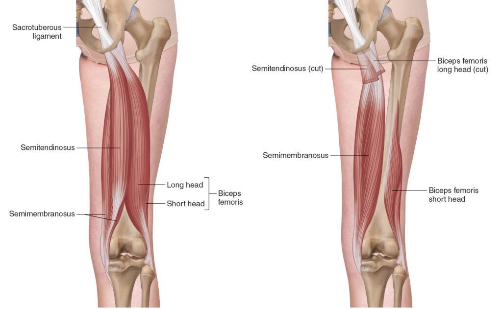
The hamstring group—biceps femoris long and short head, semitendinosus, and semimembranosus—does more than extend the hip and flex the knee. Each muscle has a unique architecture:
- semitendinosus with its long fascicles and compliant tendon
- semimembranosus with its broad pennation and large cross-sectional area
- biceps femoris long head with its short, force-oriented fibers (Kellis & Sahinis, 2021; Takeda et al., 2023).
This heterogeneity is not redundancy—it’s strategy. It allows the hamstrings to manage force differently across the gait cycle.
During the late swing of sprinting, the biceps femoris long head reaches its maximal activation while lengthening at approximately 112 % of its resting length (Chumanov et al., 2011; Andrews et al., 2025). This is where most hamstring strains occur, not because the muscle is weak but because it is forced to do two contradictory things at once—absorb and produce force.
Beyond Origins and Insertions
A muscle never acts alone. Its behavior is dictated by the joint context above and below. The hamstrings’ function depends as much on pelvic control and ankle stiffness as on their own contractile ability.
Open vs. Closed Chain Reality
In open-chain motion (like a Nordic curl), the distal segment moves freely; in closed-chain motion (like sprinting), the distal segment is fixed, and the muscle must now control the proximal segment. This reverses the apparent “action.”
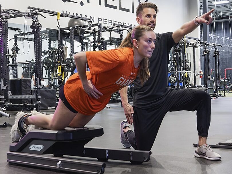
In the Nordic, the hamstrings eccentrically resist knee extension. In the sprint, they isometrically manage hip flexion torque and transmit energy into the ground (Van Hooren & Bosch, 2016a).
Open-chain drills bias local tissue capacity—building fascicle length, tendon compliance, and hypertrophy (Bourne et al., 2017). Closed-chain actions bias coordination—integrating the hamstrings with glutes, calves, and trunk to create system-level stability (Van Hooren & Bosch, 2016b). Both are essential, but their outcomes differ.
Rotary, Not Linear
Every motion in sport contains rotation—even a straight sprint.
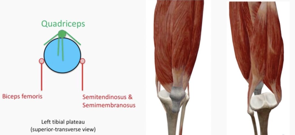
The pelvis oscillates in transverse and frontal planes; the femur spirals inward while the tibia counter-rotates. This multiplanar motion demands that muscles generate torque, not just linear force.
The semitendinosus, for example, contributes to internal rotation of the tibia during the terminal swing phase—stabilizing knee alignment while balancing the lateral pull of the biceps femoris (Yanagisawa, 2015). The interplay of these torques produces the rotational “lock-unlock” system that keeps the knee both mobile and stable.
Functional Anatomy Through the Lens of Adaptation
Fascicle Behavior: More Than a Stretch
Eccentric training lengthens fascicles, a change thought to protect against strain injury.
Research shows that athletes with fascicles shorter than 10.56 cm are four times more likely to suffer a hamstring strain (Timmins et al., 2016).
Yet the story is more nuanced.
Fascicle “lengthening” is not simply tissue elongation—it represents serial sarcomerogenesis, the addition of sarcomeres in series to distribute strain (Andrews et al., 2025). Early eccentric training increases sarcomere length; prolonged training increases sarcomere number (Pincheira et al., 2022). This adaptation reduces strain per sarcomere, allowing the muscle to tolerate long-length loading with less structural damage.
But fascicle length isn’t the whole story. Eccentric adaptations are regional: distal fascicles often adapt more than proximal ones due to nonuniform strain during training (Andrews et al., 2025). Thus, exercise selection—and even execution angle—changes which region adapts.
The Eccentric–Isometric Continuum
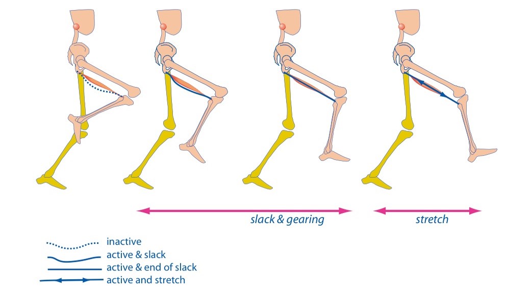
Hamstring protection isn’t only about eccentric strength; it’s about the ability to remain stiff at long lengths. Van Hooren & Bosch (2016a) argue that the hamstrings act quasi-isometrically during late swing—holding tension while the tendon lengthens and recoils.
This isometric “bracing” allows efficient elastic storage, reducing metabolic cost and mechanical risk.
In practice, this means a muscle’s ability to hold may be as valuable as its ability to lengthen. High-intensity isometrics, where the muscle resists motion while the tendon stretches, may replicate sprint demands more specifically than traditional eccentrics (Van Hooren & Bosch, 2016b).
Non-Contractile and Neural Players
Eccentric loading doesn’t just remodel fibers—it reshapes the extracellular matrix, tendon, and nervous system. Within weeks, collagen synthesis and cross-linking increase tensile capacity, while titin stiffens, improving elastic recoil (Andrews et al., 2025). Neural adaptations further enhance protective control: higher firing frequencies, reduced inhibitory reflexes, and more synchronous motor-unit recruitment (Duchateau & Enoka, 2016).
The irony? These adaptations occur across contraction types—eccentric, concentric, and isometric—provided the intensity and intent are high (Garma et al., 2007). The lesson: what matters most isn’t the mode, but the magnitude and meaning of stress.
Applying Functional Anatomy
Functional anatomy isn’t abstract—it dictates how you evaluate, cue, and progress movement.
Phase 1: Early Rehab—Reconnecting the System
In the early stages after injury, traditional rehab isolates parts: restore knee flexion, regain strength, normalize gait. Functional anatomy insists we start with coordination.
Re-establish pelvic control, trunk stiffness, and sensory feedback before adding load.
Simple drills like low-load bridge holds, heel slides, or assisted marches rebuild proprioceptive maps that reconnect muscle to movement intent.
Phase 2: Middle Phase—Reintroducing Force
As tolerance grows, the goal shifts to force management—teaching the hamstrings to accept and redirect load.
Eccentric-biased exercises such as the Nordic curl or razor curl elongate fascicles and condition neural inhibition (Bourne et al., 2017).
But supplement them with isometric and hybrid drills—Roman-chair holds, split-stance hinge isometrics, and single-leg RDL pauses—to train stiffness and rate of force development.
Phase 3: Late Phase—Reintegration and Rotation
Return-to-play success depends on rotational control.
Introduce drills that demand multiplanar timing—crossover runs, lateral accelerations, curvilinear sprints. Here, the hamstrings integrate with glutes and adductors to manage transverse-plane torques, mirroring real sport mechanics.
Phase 4: Return to Sport—Dynamic Isometrics
The final step isn’t about maximum load but maximum control. Exercises like single-leg Romanian deadlifts with perturbations, band-resisted hip drives, or “snap-back” sprints force the athlete to stabilize against unpredictable tension. These replicate the quasi-isometric reality of sprinting—the muscle holds while the system moves.
Integration with the 3P Framework
Principles
Functional anatomy brings the Coordinate Movement principle to life—it reminds us that the body doesn’t move in parts, it moves in patterns. Every muscle is a participant in a much larger conversation of timing, pressure, and direction. True proficiency isn’t built by strengthening a link; it’s built by harmonizing the chain. Strength without coordination is like volume without rhythm—loud, but out of tune.
Process
Process is where anatomy becomes behavior. Through Experiment and Exposure, we take what’s known and see how it holds up under real-world conditions—load, speed, fatigue, chaos. Every rep is a micro-experiment, a probe into how tissue, tension, and intention respond to stress. Test–retest isn’t just data collection; it’s curiosity in motion, turning the training room or weight into their own laboratory.
Plans
Plans translate anatomy and process into precision. Each variable—stance width, rotation, tempo—is a lever that shapes adaptation. A skilled therapist or coach doesn’t add noise; they manipulate signal. The art lies in amplifying what’s desirable—fluid torque, stable rotation—and dampening what distracts, like pelvic drift or tibial whip. Plans aren’t rigid scripts; they’re jazz—structured enough to guide, flexible enough to adapt.
5 Common Pitfalls of Exercise Prescription
- Over-Isolation
Treating muscles as independent units rather than contributors to force couples. - Ignoring Rotation
Linear thinking neglects transverse-plane control, where most performance gains and injuries occur. - Overemphasis on Eccentric Strength Tests
Fascicle length and Nordic strength matter, but so does the ability to coordinate timing and stiffness. - Neglecting the Environment
A muscle’s function changes with surface, footwear, and fatigue—context modifies anatomy. - Failure to Progress Across the Continuum
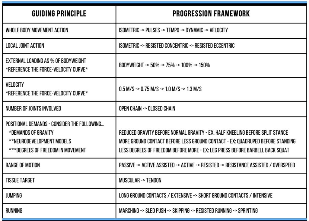
From open-chain isolation to closed-chain integration, every stage prepares the next.
Summary and Integration
Functional anatomy replaces memorization with interpretation. It’s not about where a muscle starts or ends, but how it behaves in context. Muscles are not levers; they are living sensors of load, velocity, and direction. When trained as part of an integrated system—across planes, pressures, and perturbations—they express their true purpose: coordination under constraint.
This understanding bridges the gap between rehab and performance, medicine and motion, biology and behavior. The anatomy lab gives us the map. Functional anatomy gives us the terrain.
5 Keys to Functional Anatomy in Rehab
- Measure What Matters. Use embedded testing—force, timing, symmetry—to quantify true function.
- Think in Systems, Not Segments. Every muscle action depends on its neighbors.
- Respect Rotation. Torque drives coordination.
- Train Stiffness as Much as Strength. Isometric intent builds control.
- Progress from Isolation to Integration. Move from open- to closed-chain control.
Watch More Like This
Read More Like This
Related Podcasts
References
- Andrews, M. H., Shield, A. J., Lichtwark, G. A., & Pincheira, P. A. (2025). Hamstring injury mechanisms and eccentric training-induced muscle adaptations: Current insights and future directions. Sports Medicine.
- Bourne, M. N., Duhig, S. J., Timmins, R. G., Opar, D. A., & Shield, A. J. (2017). Impact of the Nordic hamstring and hip extension exercises on hamstring architecture and morphology. British Journal of Sports Medicine, 51(5), 469–477.
- Chumanov, E. S., Heiderscheit, B. C., & Thelen, D. G. (2011). Hamstring musculotendon dynamics during stance and swing phases of high-speed running. Medicine & Science in Sports & Exercise, 43(3), 525–532.
- Garma, T., et al. (2007). Similar acute molecular responses to isometric, lengthening, or shortening resistance exercise. Journal of Applied Physiology, 102, 135–143.
- Kellis, E. (2018). Intra- and inter-muscular variations in hamstring architecture and mechanics. Sports Medicine, 48, 2271–2283.
- Kellis, E., & Sahinis, C. (2021). Effect of knee joint angle on individual hamstring morphology. Journal of Electromyography and Kinesiology, 62, 102619.
- Pincheira, P. A., et al. (2022). Biceps femoris long head sarcomere and fascicle length adaptations after 3 weeks of eccentric training. Journal of Sport and Health Science, 11, 43–49.
- Stępień, K., et al. (2018). Anatomy of proximal attachment, course, and innervation of hamstring muscles. Knee Surgery, Sports Traumatology, Arthroscopy, 27, 673–684.
- Takeda, K., et al. (2023). Unique morphological architecture of the hamstring muscles. Journal of Anatomy, 243, 284–296.
- Timmins, R. G., et al. (2016). Short biceps femoris fascicles and eccentric knee-flexor weakness increase risk of hamstring injury. British Journal of Sports Medicine, 50(24), 1524–1535.
- Van Hooren, B., & Bosch, F. (2016a). Is there really an eccentric action of the hamstrings during the swing phase of high-speed running? Part I. Journal of Sports Sciences, 34, 2313–2321.
